A 3D Printed Clip-on Turns Your Phone into a Microscope

 3D Printing, Smart Phones  Comments Off on A 3D Printed Clip-on Turns Your Phone into a Microscope
Mar 062018
 

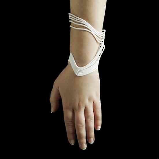
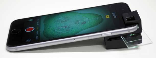
Anyone, whether you are a scientist or are just curious, can now turn the humble smartphone into a fully functional microscope. Researchers behind the development of the tool have made the 3D files publicly available. So if you have a 3D printer, you can create the microscope and soon get started on examining various samples.

This isn’t the first attempt at turning a smartphone into a microscope, but it shown the most promise. Developed by researchers at the ARC Centre of Excellence for Nanoscale BioPhotonics of the RMIT University in Australia, the clip-on magnifies up to 1/200th of a millimeter. It’s also different from previous versions since it doesn’t use external light or power sources.

smartphone microscope


Previous attempts at creating a smartphone microscope have made use of LEDs and power sources, but Australian researchers tapped into resources available on the mobile device itself. Dr Antony Orth, lead developer on the project, said that their mobile phone microscope uses the “integrated illumination” available in almost all smartphone cameras.

The clip-on was designed with internal illumination tunnels that guide the light from the flash of the camera to light the sample from behind. With this, Orth and fellow researchers have overcome an issue with other phone-based microscopes: the elimination of additional illumination optics.

With a smartphone microscope, the cost of and complexity of assembly is significantly reduced. There is only one simple assembly step required to get the device ready to go. Orth says that there is a lot of potential for the clip-on to be used as a scientific tool.

He adds that the 3D printed clip-on can be taken along for onsite or remote-area monitoring. Traditional equipment for such activities tend to be bulky, which makes them impractical. So the availability of a much smaller device that can perform the same task makes outdoor studies more convenient.

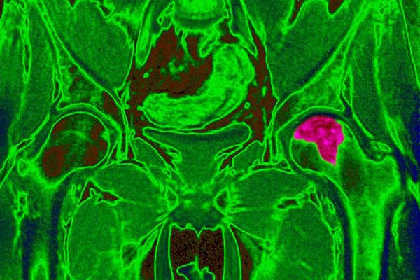
Capable of visualizing specimens as small as 1/200th of a millimeter, this clip-on device can be used to examine animal and plant cells, blood cells, cell nuclei, and microscopic organisms, among others.

The team behind the mobile device microscope expects it to be used for tasks such as testing the cleanliness of water, detecting disease, and checking blood samples for the presence of parasites.

Orth and his colleagues have tested the microscope in several areas. They have successfully visualized samples such as zooplankton and semen from live cattle. With the files available to the public, just about anyone with access to a 3D printer can start being curious about the small things.


References

http://cnbp.org.au/online-tools – 3D printing files

https://www.engadget.com/2018/02/20/3d-printed-smartphone-microscope-is-good-enough-for-scientists/


Swedish Scientists Use 3d Bioprinters to Generate Human Cartilage

 3D Printing  Comments Off on Swedish Scientists Use 3d Bioprinters to Generate Human Cartilage
May 162017
 

Lead Swedish researcher, Stina Simonsson, is in high spirits as she claims that her research team, created from the collaboration between scientists from Chalmers University and Sahlgrenska Academy from Belgium, has made a breakthrough in the field of medicine. This was after they were able to produce 3D-printed stem cells from cartilage cells extracted from knee surgery patients.

According to Simonsson, while the process to naturally differentiate stem cells into cartilage is not complicated, they were the first ones to successfully generate artificial cartilage, using real stem cells, which can be used for osteoarthritis treatment and damaged cartilage tissue repair. They made use of 3D bioprinting, also referred to as additive manufacturing, a process applied in regenerative medicine to address the issue of the rise in demand for engineered organs and tissues for transplantation on patients.

The process, posited by Simonsson, did not even require subjecting animals for testing and the good news being the stem cells were able to survive the printing process.


How does 3D bioprinting work?

After cartilage cells are harvested from patients who have undergone knee surgery, these cells are then transformed back into “pluripotent” stem cells. These stem cells are called the master cells since they have the potential to form all other cell types.

After being reverted, these cells are then covered in nano-cellulose material and bio-printed. The nano-cellulose compound is instrumental in the survival of these cells during printing. When they survive, growth stimulants are used to aid in cell differentiation and multiplication. Consequently, artificial cartilage will be formed.

However, the research team also disclosed one challenge they need to deal with. According to them, before patients can receive 3D bioprinted cartilage implants, further exploration should be done to find the perfect cellulose material that can be broken down properly and accepted by the body to ensure what remains is endogenous cartilage.

If this technique is applied, they also warned that more live stem cells will be needed. At the moment, the cellulose they have used is believed to be not perfectly suited for the human body.

The process of 3D bioprinting has already been in existence in recent years, with medical researchers from other parts of the world, including the U.S and Canada, trying this technology. Four years ago, scientists from Cornell University were able to generate human ears from cells harvested from a cow and 3D bioprinted. Conversely, Princeton University also reached a breakthrough when researchers used a 3D bioprinter to grow ears. They even claimed that these ears are able to receive frequencies from a very far range, outdoing the hearing capabilities of humans.

In Toronto Canada, the collaboration between the researchers from the University of Toronto and the Ross Tilley Burn Centre, resulted in the development of a process to mimic the appearance and qualities of human skin. This can be used for burn patients in the future, according to Dr. Marc Jeschke.

Meanwhile, despite the need for exploring new cellulose material, the hopes of researchers remain high. They believe that in the time, they can achieve their goals since the first steps have been taken and results are positive.


References

http://www.cbsnews.com/news/scientists-hit-milestone-in-successful-3d-printing-of-cartilage/

https://cosmosmagazine.com/biology/stem-cells-survive-3d-printing-to-produce-cartilage


Madeline Gannon and Tactum: 3D Design and Printing for All

 3D Printing  Comments Off on Madeline Gannon and Tactum: 3D Design and Printing for All
Sep 152015
 

Unlike regular printing which almost all of us know how to do without having to scratch our eyes out, 3D design and printing is in a league of its own. Basically, it’s more tailored towards those who are more tech savvy and actually love creating three-dimensional objects. However, Madeline Gannon – a researcher and teacher at the Carnegie Mellon University School of Architecture and PhD candidate in Computational Design – wants to change that. She wants to unleash the designer that is hiding in all of us.

Gone are the days when 3D printers used to be luxury machines. Although there are still rather expensive models these days, technology has advanced so much that anyone with a few hundred dollars to spare can get their hands on a 3D printer. However, as Gannon notes, not everyone can just create original 3D objects.


Enter Tactum

In order to give creative power to ordinary 3D printer owners, Gannon developed a system called Tactum. It’s an innovative software system that gives users the ability to create their own designs for 3D printers by just touching a projected image.

Essentially, one would just have to rub, poke or use any other hand gestures on a projected image which will then become their 3D printed object. Through this process, people can instantly see their object change shape in response to the touches.

The first series of 3D objects Gannon designed made use of a surface that is very much accessible: the human body.

Together with a companion project called Reverb which helps convert user-created designs into printable meshes, Gannon has created bracelets and necklaces with wide-ranging designs, including smooth landscapes and intricate textures.


Further Uses

Tactum’s use in creating fashionable items is just the beginning. The system really proves itself when used in the creation of functional pieces like the custom watchband Gannon designed for a Motorola Moto 360 smartwatch.

Gannon plans to use Tactum for customizing prosthetics and other wearable medical devices. Can you just imagine how better it would be for patients to have a truly customized device? They can collaborate with doctors and a Tactum technician in real time, providing feedback regarding the fit and feel of the device.

Tactum has the potential to produce 3D objects at a rate that is much quicker and at even lower cost. As such, doctors and patients can continually adjust prosthetic limb as they see fit, and also giving the patient a certain degree of personal expression.


The Journey to Maker

Gannon’s path to Maker can be traced back to her trips to museums as a high school student. For her, the buildings interested her more than the exhibits themselves. She realized then that she wanted something to do with architecture.

However, during her last year of architecture school, she experienced the limits of the human-computer interface. In other words, the computer couldn’t produce the ideas from her head. As a result, Gannon plunged herself into computer science.

Fast forward to the present and Gannon now heads MADLAB.CC, a design collective that explores computational approaches to architecture craft and interaction all aimed at exploring the “edges of digital creativity.”