Kevin Lepton

Gene-Editing Medicine Breakthrough

 Future Medical Technology  Comments Off on Gene-Editing Medicine Breakthrough
May 072018
 

Human Gene Editing


For years, the concept of gene-editing has been consigned to fictional movies and grandiose works of writing. However, it appears that with a major breakthrough that has taken place recently, we might be closer to a breakthrough than we would ever have expected.

The concept is simple: a small biomachine will enter the human body. Then, it looks to find out where defective gene sequences begin and end. Then, it will edit the defect and implant the ‘right’ information instead, with immense accuracy. If it sounds like something for next century, you’ll be pleased to know that this could arrive sooner rather than later.

This form of gene editing has been under development at the University of Alberta. Researchers have produced an exceptional new study that believes the reality is much closer than one would expect.

According to Basil Hubbard, the Canada Research Chair in Molecular Therapeutics and an assistant professor at the University, progress is very impressive. “We’ve discovered a way to greatly improve the accuracy of gene-editing technology by replacing the natural guide molecule it uses with a synthetic one called a bridged nucleic acid, or BNA,” he said.

At the moment, the University has pushed for a patent to help make sure it stays locked as their discovery. Also, they are looking for pharmaceutical industry experts to partner with them to turn this into a genuine therapeutic solution.

Ever since the discovery of the CRISPR/Cas9 system in bacteria gene-editing has become a popular topic of discussion. This form of solution is used to help protect against natural predators known as bacteriophages. Hubbard explained it as allowing “bacteria to store information about previous infections and then use it to seek out and destroy the DNA of new invaders by cutting it.”

The hope is that we ca use this to help cut a specific DNA sequence within a human too. This would allow for gene manipulation. However, the main issue at the moment is the lack of specification that is present; at the moment, it’s cutting similar – but still wrong – genes during the process.

While it’s only making mistakes in around 1% of cases, that’s still too much, as Hubbard explained. “However, given that there are trillions of cells in the human body, even one percentage off is quite significant, especially because gene editing is permanent. One wrong cut and a patient could end up with a serious condition like cancer.”

Although there’s still a large amount of hurdles to be overcome with this particular system, gene-editing just became more realistic. There’s a lot of changes to come, including how to effectively produce it within a human body. However, we are now many more steps closer to making the power of gene-editing a genuine medical solution.

If you would like to know more about this, then check out the Nature Communications journal where you can find the full study. It’s a truly special step towards a safer, healthier future for all.
3d Printed Models of Heart Help Surgeons Prepare for Surgery

 3D Printing, Future Medical Technology  Comments Off on 3d Printed Models of Heart Help Surgeons Prepare for Surgery
Apr 052018
 

3d Printed Heart


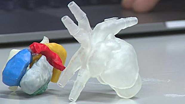
As one of the most difficult medical and surgical processes, the human heart transplant is a major challenge. The news, then, that a team of Wisconsin surgeons are working with a 3D printing firm to change that is entirely good news. These surgical experts are teaming up with engineering teams to get access to 3D-printed heart models. These models will be used to help a surgeon to prepare on a direct copy of their upcoming patient’s heart.

Aimed at helping to make pediatric heart surgery more likely to be a success, this is a positive move. The aim is to help make sure that there is a more specific accuracy with regards to the surgery outcome.

Speaking about the idea was Dr. Petros Anagnostopulous, the Chief Paediatric Cardiothoracic Surgeon at the American Family Children’s Hospital. A member of the University of Wisconsin Health faculty, too, he’s a major advocate of the idea. Speaking to WSAW.tv, he was very clear about the benefits this could bring.

Big Advantage

“The big advantage of the 3D printing is that you can recreate the heart. It’s the closest I’ve ever seen, some model — to the actual thing,” he said.

With the aim being to work on a printed version of the heart, the hope is that surgeons can vastly improve upon their accuracy for a specific patent. Already, one patient has benefited from this service – Joseph Oehlof, 6. Joseph was in need of a transplant after suffering from a rare heart condition.

Taken to the American Family Children’s Hospital for care, Dr. Anagnostopulous was able to carry out the treatment. He used a 3D model of the heart to help better prepare him for the challenging surgical scenario to come.

“There’s a lot of ability to see the relationship of the different parts of the heart as they are in real time. The 3D model prepares you better. The other thing it can do; it prepares your whole team better.” Dr. Anagnostopulous said.

Team Work

The developers of this unique technology are at the University of Wisconsin, too. Headed up by Alejandro Roldan Alzate, the Engineering Professor believes this is a major step forward for medical science. “When the surgeon can interact with a 3D printed heart before surgery, that helps a lot,” Alejandro said.

“What we have at UW as a benefit is that the medical school and the engineering school are just a bus ride away. All of the advances we have in engineering can be translated immediately in to the hospital.”

With the aim being to eventually take this into orthopaedic surgical practices, too, this could go very far. The hope is that, with the success of this practice, that other challenging parts of the human body will soon be fully accessible in preview form prior to the surgery taking place.


A 3D Printed Clip-on Turns Your Phone into a Microscope

 3D Printing, Smart Phones  Comments Off on A 3D Printed Clip-on Turns Your Phone into a Microscope
Mar 062018
 

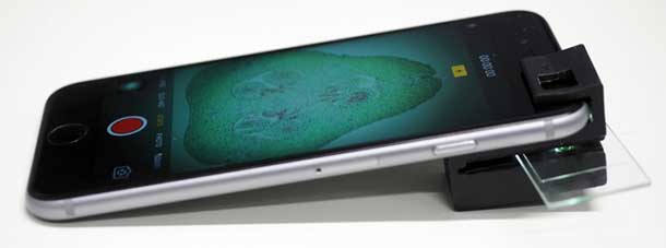
Anyone, whether you are a scientist or are just curious, can now turn the humble smartphone into a fully functional microscope. Researchers behind the development of the tool have made the 3D files publicly available. So if you have a 3D printer, you can create the microscope and soon get started on examining various samples.

This isn’t the first attempt at turning a smartphone into a microscope, but it shown the most promise. Developed by researchers at the ARC Centre of Excellence for Nanoscale BioPhotonics of the RMIT University in Australia, the clip-on magnifies up to 1/200th of a millimeter. It’s also different from previous versions since it doesn’t use external light or power sources.

smartphone microscope


Previous attempts at creating a smartphone microscope have made use of LEDs and power sources, but Australian researchers tapped into resources available on the mobile device itself. Dr Antony Orth, lead developer on the project, said that their mobile phone microscope uses the “integrated illumination” available in almost all smartphone cameras.

The clip-on was designed with internal illumination tunnels that guide the light from the flash of the camera to light the sample from behind. With this, Orth and fellow researchers have overcome an issue with other phone-based microscopes: the elimination of additional illumination optics.

With a smartphone microscope, the cost of and complexity of assembly is significantly reduced. There is only one simple assembly step required to get the device ready to go. Orth says that there is a lot of potential for the clip-on to be used as a scientific tool.

He adds that the 3D printed clip-on can be taken along for onsite or remote-area monitoring. Traditional equipment for such activities tend to be bulky, which makes them impractical. So the availability of a much smaller device that can perform the same task makes outdoor studies more convenient.

Capable of visualizing specimens as small as 1/200th of a millimeter, this clip-on device can be used to examine animal and plant cells, blood cells, cell nuclei, and microscopic organisms, among others.

The team behind the mobile device microscope expects it to be used for tasks such as testing the cleanliness of water, detecting disease, and checking blood samples for the presence of parasites.

Orth and his colleagues have tested the microscope in several areas. They have successfully visualized samples such as zooplankton and semen from live cattle. With the files available to the public, just about anyone with access to a 3D printer can start being curious about the small things.


References

http://cnbp.org.au/online-tools – 3D printing files

https://www.engadget.com/2018/02/20/3d-printed-smartphone-microscope-is-good-enough-for-scientists/